Extended Depth-of-Field Digital Microscope SystemSDM-1000 Series
For electronic components, precision parts, metal materials and complex surface samples, SDM-1000 combines overview positioning, full-frame clear observation, local magnification, 2D measurement and 3D morphology-assisted analysis in one microscopy workflow.
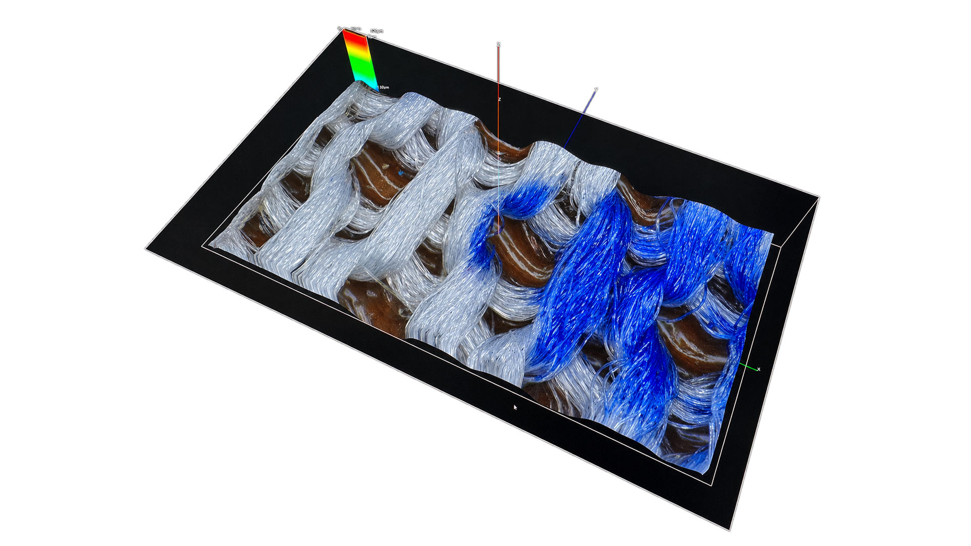
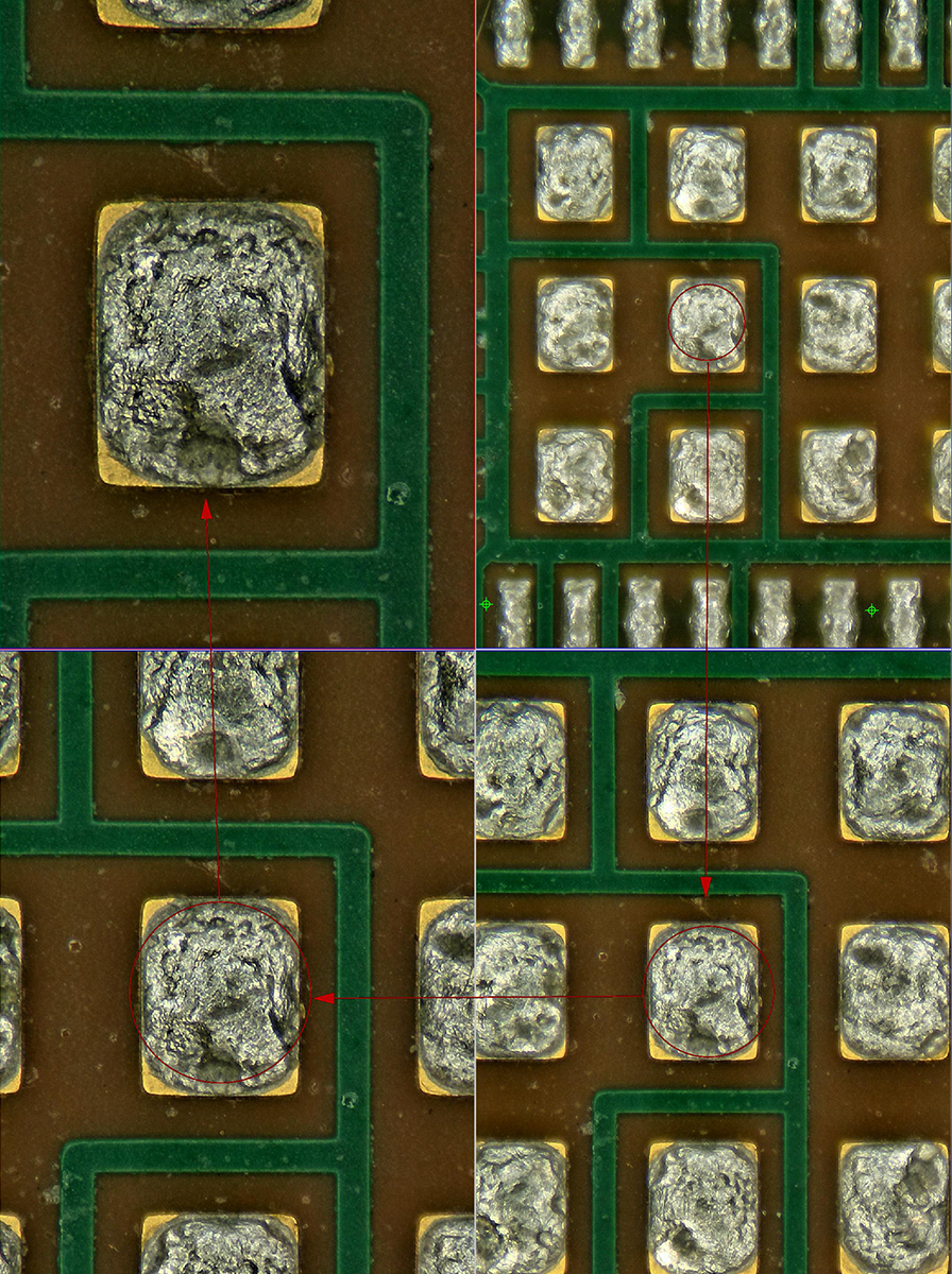
The SDM-1000 Series is an Extended Depth-of-Field Digital Microscope System for industrial surface observation, defect inspection and microscopic analysis. For samples with height differences, concave-convex structures or complex textures, the system collects images from different focal planes and synthesizes them into one extended-depth image, keeping areas at different heights clear at the same time.
Users can start with sample overview positioning, then continue with local magnification, multi-directional lighting observation, 2D dimensional measurement and 3D morphology-assisted analysis. Image acquisition, observation, measurement and result output stay in the same workflow, reducing the burden of repeated focusing, device switching and sample repositioning.
02 / Extended depth-of-field synthesis
From the highest point to the lowest point, it remains clear.
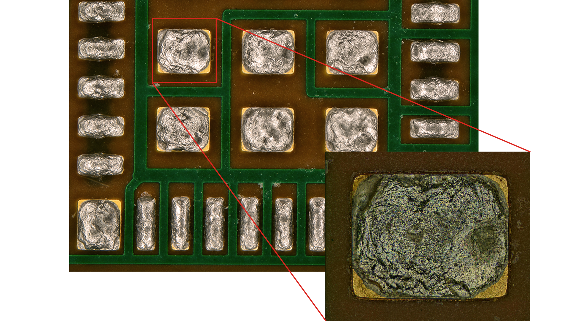
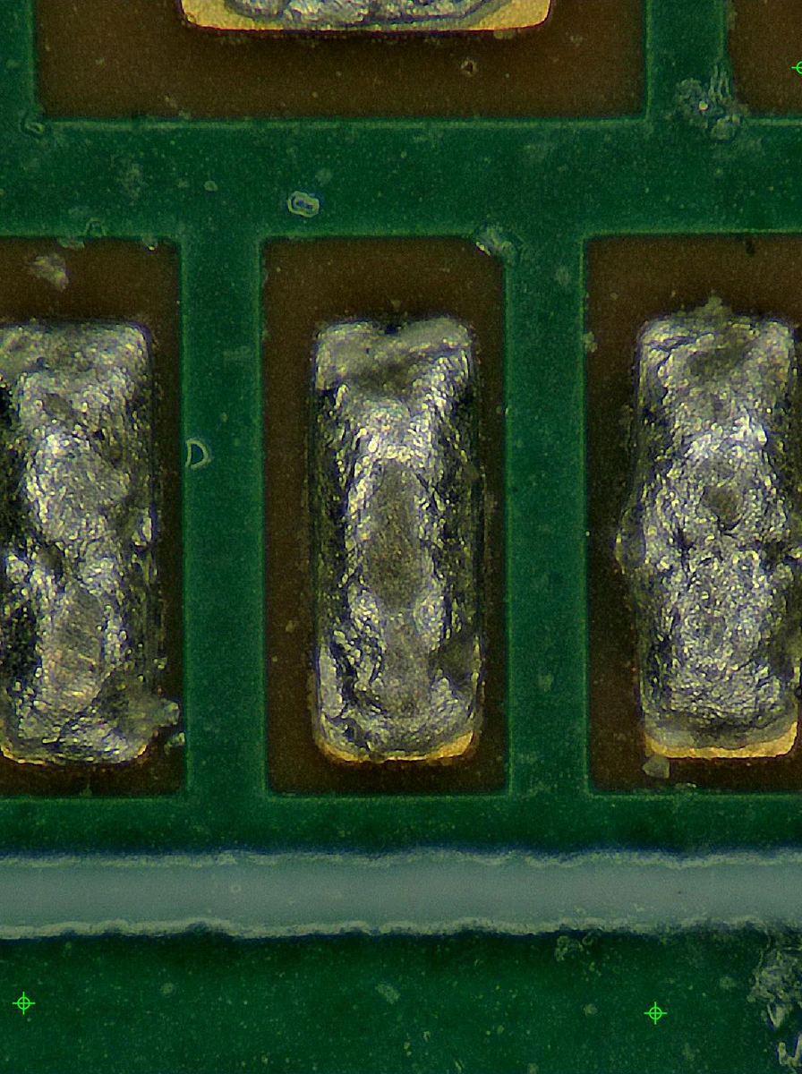
The system collects clear information from different height positions of the sample and combines it into a full-frame focused image. For solder joints, fractures, burrs, tools, connectors and complex surfaces with obvious height differences, users can view overall structure and local detail at the same time without repeatedly adjusting focus by hand.
03 / Continuous autofocus
Move and observe, the focus continues to follow.
When the stage moves or the observation position changes, the system can continuously adjust the focus according to the height change of the sample surface, reducing the need to re-find the focal plane. Users can complete sample inspection, position switching and key area review more consistently.
04 / Multi-directional lighting and DPC
Change the lighting direction, and the surface information will appear.
The system collects illumination images from multiple directions and combines them with differential phase contrast processing, turning tiny bumps, edges and texture changes into clearer light-dark relationships. Shadow direction can still be adjusted after capture, helping users understand the surface structure from different angles.
05 / Overall positioning and large-area stitching
Look at the overall picture first, and then go into the details that need to be reviewed.
The system can establish an overview of the sample through measurement navigation, select the target area in the overview image, and move the stage to the corresponding position. For samples beyond a single field of view, more complete observation results can be obtained through multi-field image stitching, and the system can continue to return to the local area for magnified inspection.
06 / Observation, measurement and result output
From seeing problems to leaving reviewable results.
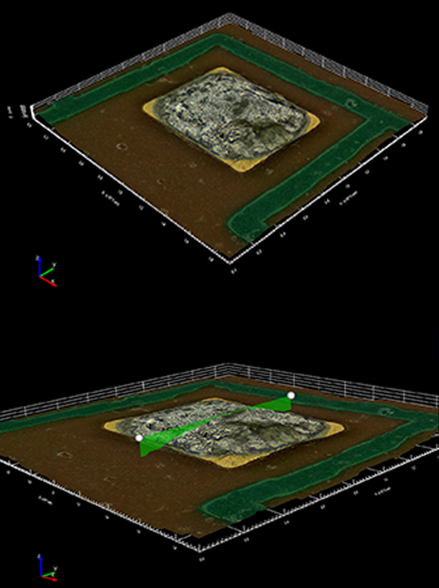
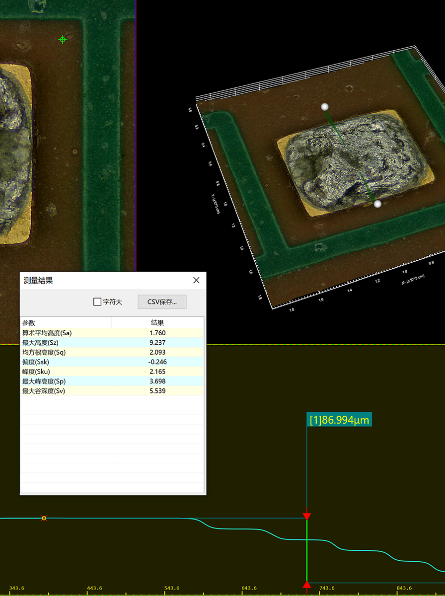
After image observation, users can continue with 2D measurements such as length, distance, angle and area, and conduct 3D morphology-assisted analysis according to the configuration. Observation images, annotations, measurement results and reports can be saved and output in the same workflow for R&D records, quality review and problem communication.
Ordinary microscopic observation often keeps only one height position in focus. For solder joints, fractures, burrs, threads and other samples with obvious height differences, the SDM-1000 Series collects images from multiple focal planes and synthesizes them so areas at different heights can be seen clearly in one image.
02 / Multi-directional lighting
Same surface, understood from different directions.
Samples that are reflective, have low contrast or have subtle textures often require changing the lighting direction to reveal key features. The system can collect lighting information from multiple directions and combine it with DPC processing to enhance the light and dark differences between surface unevenness, edges and textures.
03 / Navigation and stitching
From the entire sample, quickly return to the target location.
For samples that exceed a single field of view, the system can create an overview through multi-field image stitching, then select the area that needs review from the overview map. The stage moves automatically to the corresponding position, keeping overall positioning and local high-magnification observation connected.
04 / Measurement and analysis
After observation, continue into measurement and records.
The system connects image observation, dimensional measurement, 3D morphology-assisted analysis and result output in the same operating process. Users can mark and analyze defect locations, structural dimensions and surface changes, then use images and results for quality judgment, R&D records and communication review.
Model and configuration
One microscopy workflow, two observation scales
SDM-1100 and SDM-1200 share extended depth-of-field synthesis, multi-directional illumination, DPC, image stitching, 2D measurement and result-output capabilities. The choice depends on whether the task prioritizes coverage efficiency for daily industrial observation or high-magnification review at a smaller scale.
SDM-1100: Medium-to-low magnification model.Medium-to-low magnification model
SDM-1100
Covers a wider range of daily industrial observation and routine analysis tasks
SDM-1100 is designed for conventional industrial microscopic observation, overall positioning and local detail confirmation. Its 20×-2000× magnification range covers the observation process from sample overview to local magnification, and suits common tasks such as solder joints, burrs, fractures, machining textures, connectors and electronic component surfaces.
For users who value sample adaptability, observation efficiency and daily analysis workflows, SDM-1100 provides a balanced magnification configuration while retaining the series' extended depth-of-field synthesis, multi-directional illumination, DPC, 2D measurement and image result-output capabilities.
SDM-1200: Medium to high magnification upgrade.Medium-to-high magnification upgrade
SDM-1200
Extends observation from routine details to smaller scales
While retaining 20× initial observation, SDM-1200 extends the upper magnification limit to 6500×, making it more suitable for small defects, subtle edges, material textures and high-magnification review tasks. Users can complete overall positioning first, then move gradually into smaller scales without repeatedly switching observation devices.
Rather than simply adding software functions, it concentrates the series' observation capability on high magnification and fine detail presentation, making it suitable for R&D, failure analysis and precision quality verification that require richer surface information.
The following content shows the currently confirmed model positioning and magnification range of SDM-1100 and SDM-1200. Other optical, platform, camera and environmental parameters are subject to the official product brochure and project configuration.
Parameter
SDM-1100
SDM-1200
Model positioning
Medium-to-low magnification model
Medium-to-high magnification upgrade
Magnification range
20×-2000×
20×-6500×
Observation Focus
Overall positioning, routine detail observation and daily analysis
Tiny structure, high magnification details and precision review
Observation of PCB, solder joints, connectors, electronic components, packaging structures and surface defects.
Precision Machining
Tool, burr, fracture, processing texture, thread and surface inspection of precision parts.
Materials Research
Observation of surface structure and texture of metals, ceramics, composite materials, coatings and materials.
Automotive and New Energy
Inspection of battery materials, electrode sheets, seals, gears, bearings and automotive electronic components.
Let the samples speak first.
Submit the sample type, observation magnification and issues to confirm. An engineer will help determine the more suitable model and configuration based on the actual observation task.