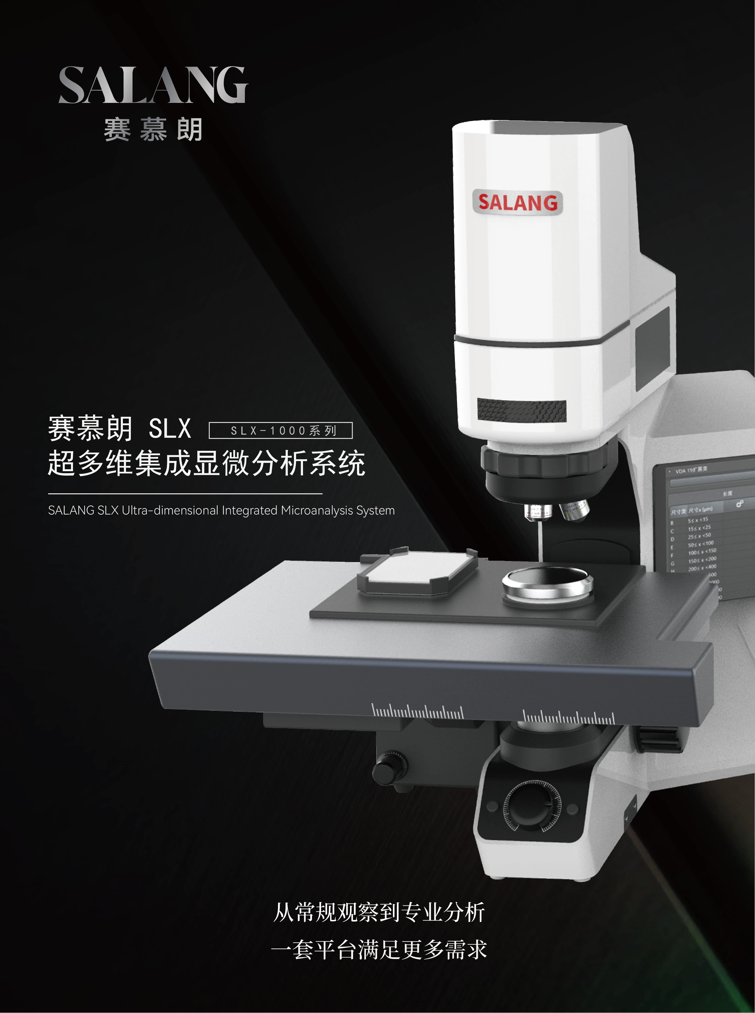
Silex Integrated Multidimensional Microscopy SystemSLX-1000 Series
Organize bio-fluorescence, metallography, professional polarization, infrared, Raman and 3D measurement tasks on the same scalable microscopy platform. Select the measuring head, objective, illumination and analysis module around the sample and task, then move from routine observation to professional analysis in one workflow.
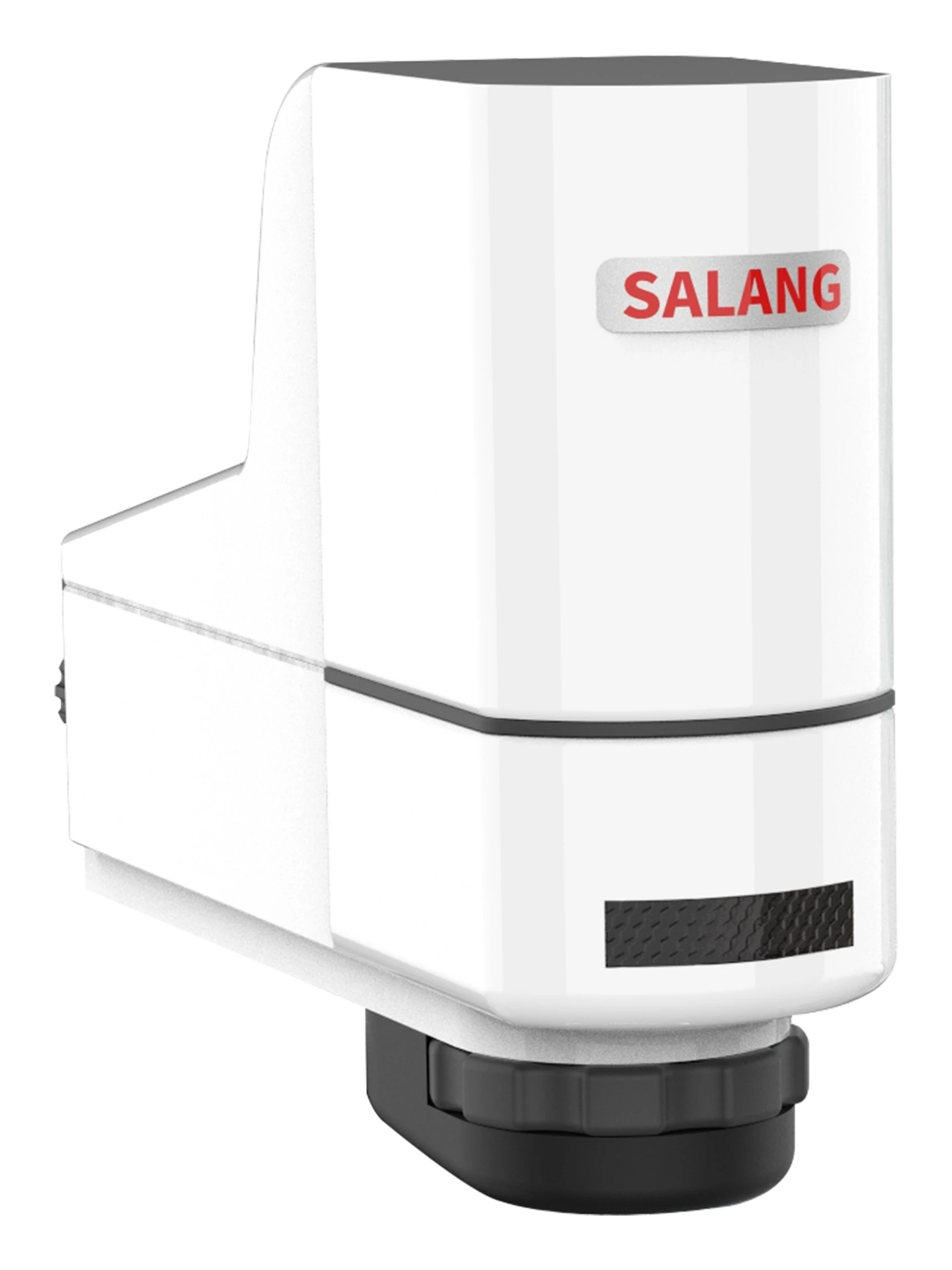
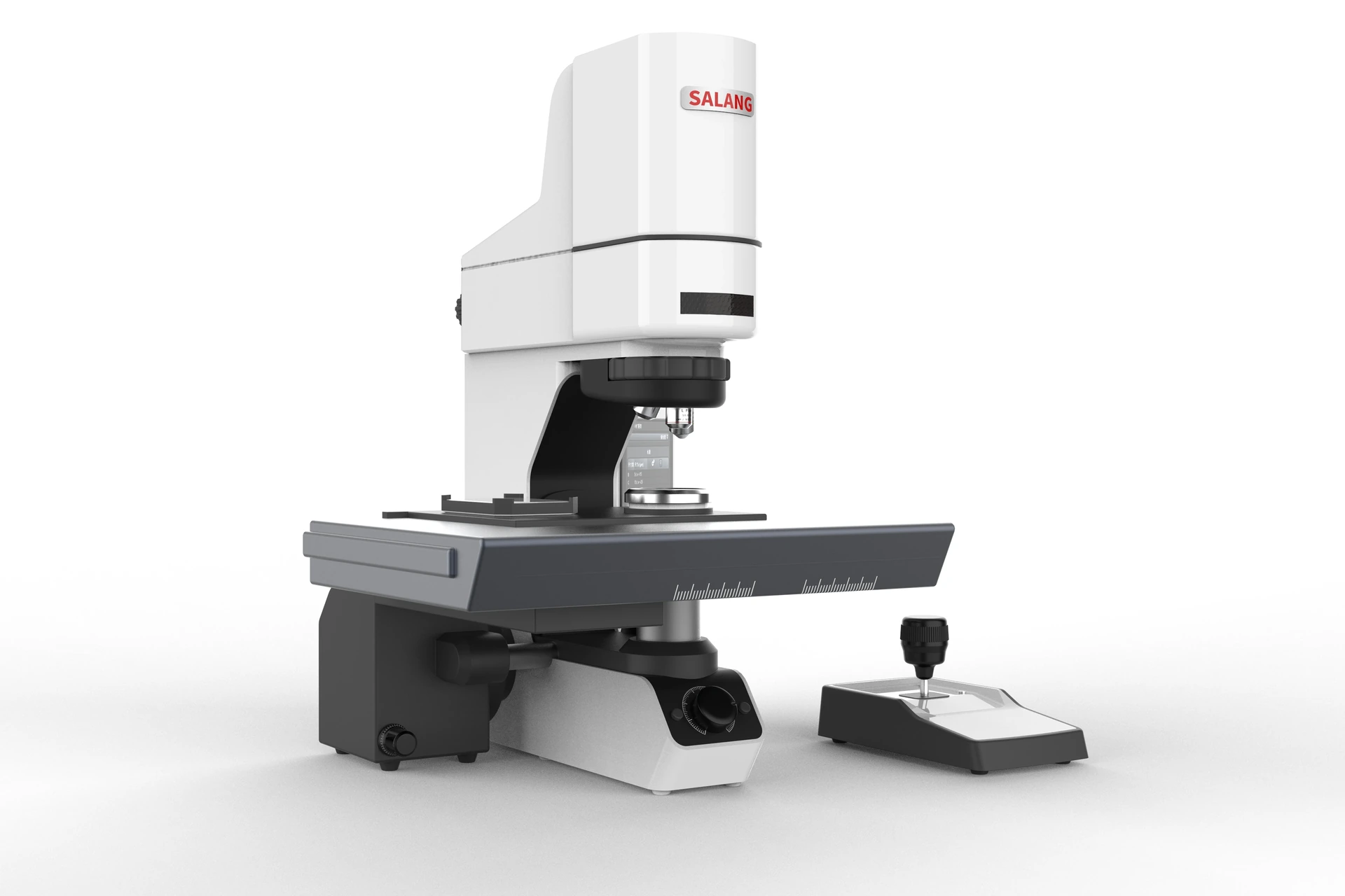
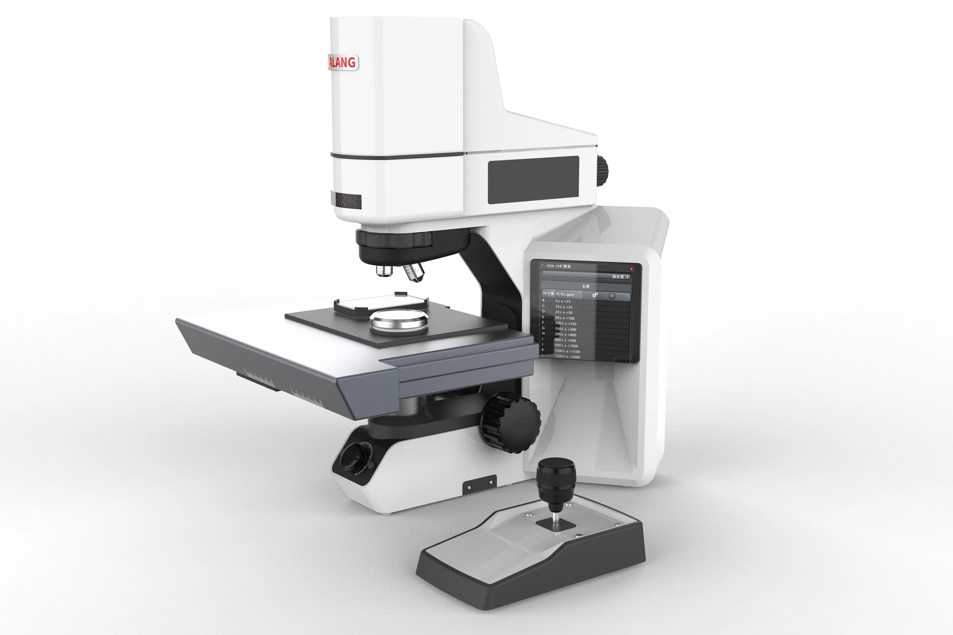
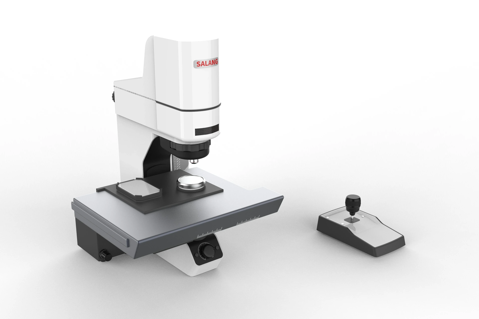
The SLX-1000 Series is an integrated microscopy platform built around modular measurement heads, an infinity-corrected optical system and unified analysis software. The system can be configured for bio-fluorescence, metallography, professional polarization and 3D measurement, with infrared, Raman and other observation capabilities added according to actual tasks.
For laboratories, R&D departments and quality centers that handle multiple samples and analysis tasks, users do not need separate hosts for each observation method. Samples can complete overview positioning, mode switching, local observation, dimensional measurement and analysis-result recording on the same platform, reducing time and data differences caused by sample transfer, repeated clamping and repositioning.
SLX-1000 Bio-fluorescence directionSLX-1100 Metallographic analysis direction
SLX-1200 Professional polarization direction3D measurement and software workflow
For the same sample, switch to different observation dimensions.
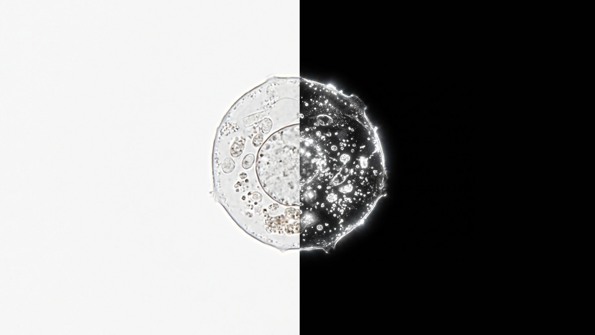
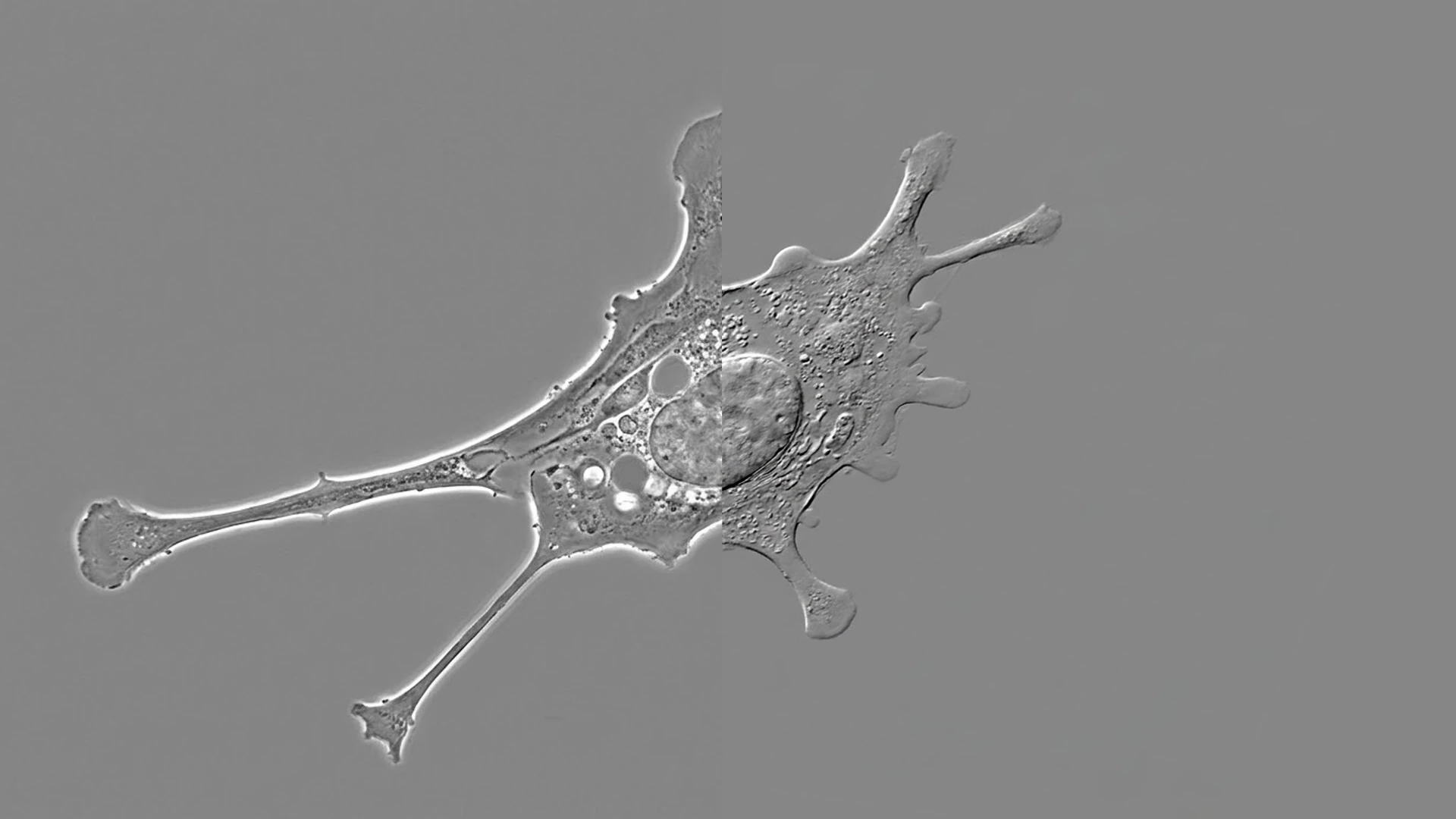
Depending on the measuring head, objective and illumination configuration, the system can observe through brightfield, darkfield, phase contrast, differential interference contrast (DIC), polarization, fluorescence and infrared modes. Each mode enhances different information, such as surface morphology, edges, tissue, fluorescence signals, material orientation or internal structure, helping users understand the same sample from different angles.
03 / Cross-scale observation and large-field stitching
Build the overview first, then move into the areas that need analysis.
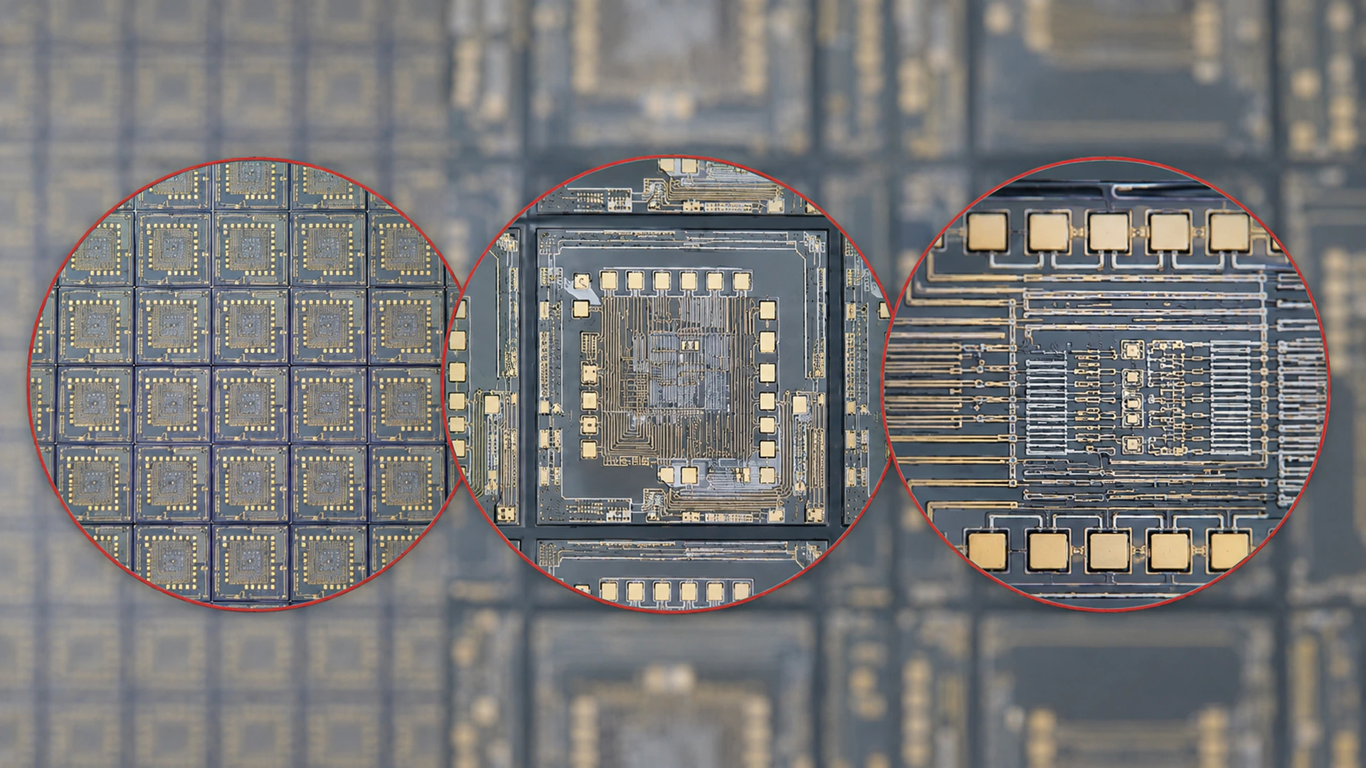
For samples beyond a single field of view, the system can build a sample overview through multi-field acquisition and let users select the location that needs further observation from the overview map. Users can move from low-magnification positioning to high-magnification local analysis, keeping large-area archiving, area review and multi-mode observation continuous.
04 / Observation, Measurement and Analysis
Capture images and turn them into analyzable data.
After microscopic observation, users can continue with 2D measurements such as length, distance, angle and area, then perform 3D morphology, surface-structure or professional analysis according to the selected measuring head and system configuration. Images, annotations, measurement results and analysis records can be saved and exported in the same software process.
Microscopic imageSelection and labeling2D dimensions3D morphologyAnalysis resultsReport output
05 / Professional capabilities expanded by task
One platform, from precision manufacturing to life science.
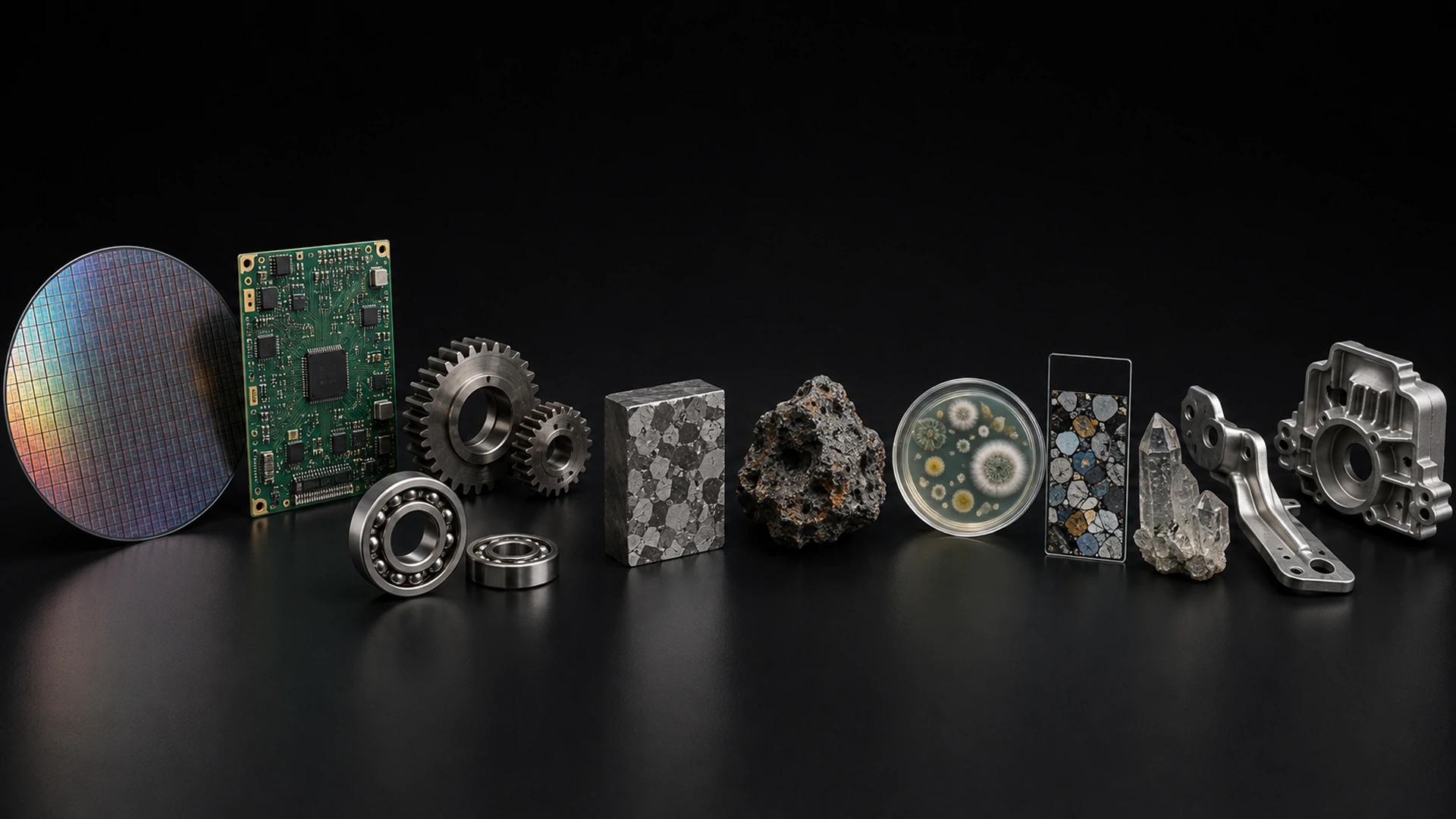
With modular optics and analysis capability, the Silex Integrated Multidimensional Microscopy System can be configured for different samples and inspection tasks. It supports applications across life science, semiconductors and electronics, materials and metallurgy, automotive and new energy, geology and minerals, and precision manufacturing—helping teams complete more microscopic tasks on one platform, from research observation to quality inspection and failure analysis.
Current industry sample focus area
01 / Multi-mode observation
Switch optical paths, reveal more dimensions of the sample.
A single observation method often shows only part of a sample. With the appropriate measuring head and configuration, SLX can switch between brightfield, darkfield, DIC, polarized light, fluorescence and infrared observation to present surface morphology, edges, tissue, fluorescence signals and internal structure more clearly.
02 / Overview and detail
Change magnification, and settings follow automatically.
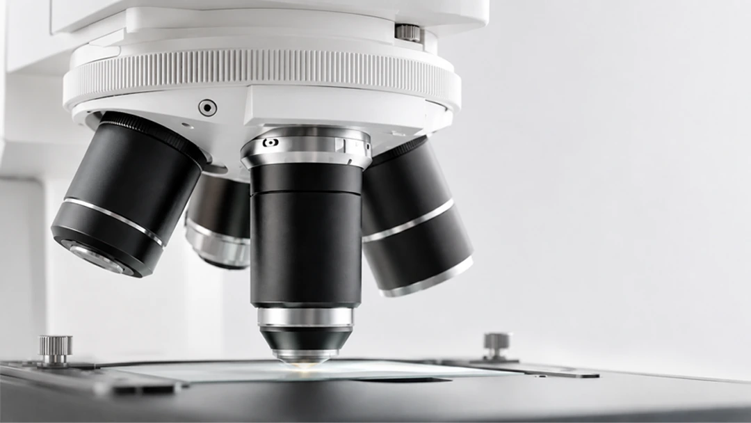
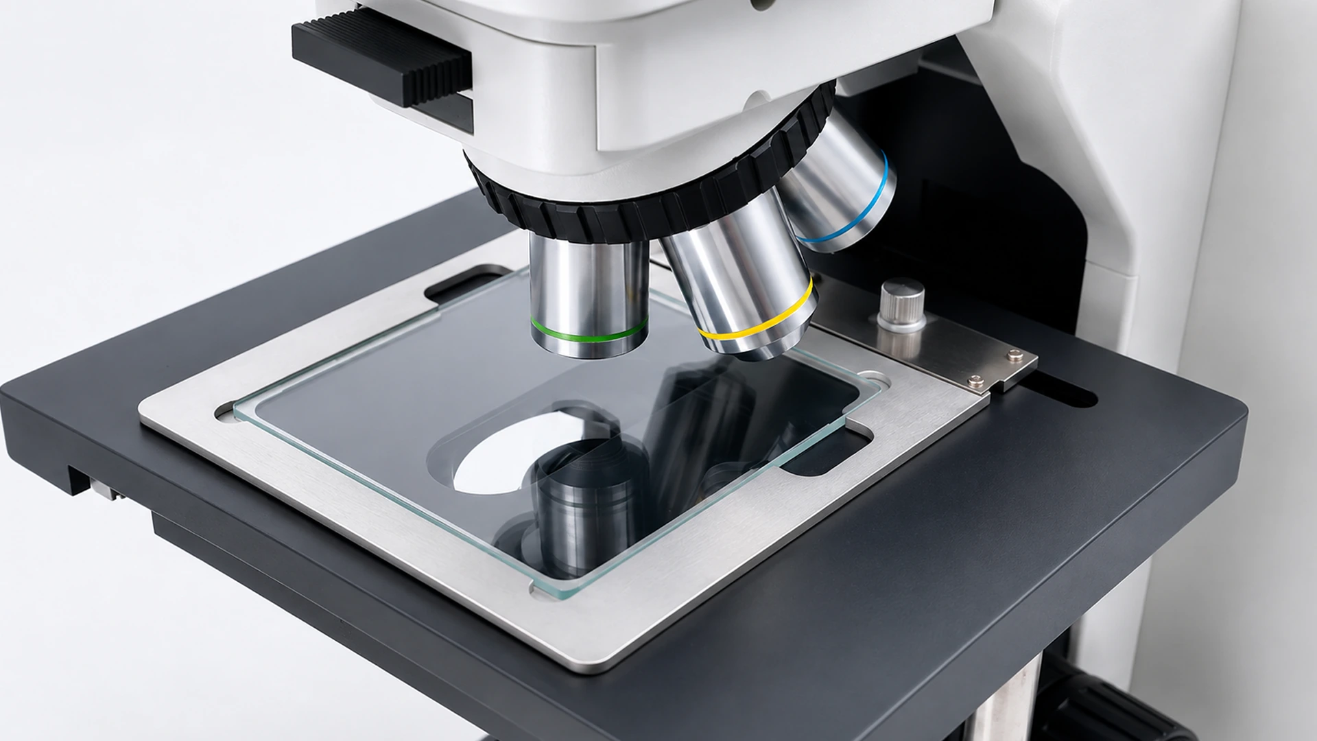
The encoded objective changer links microscope hardware with image-analysis software. When objectives are changed, the system identifies the active magnification and calls the corresponding calibration settings while remembering illumination brightness for each objective. This reduces repeated setup and measurement error during magnification changes.
03 / Measurement and Analysis
From transparent materials to metal workpieces, one platform observes them all.
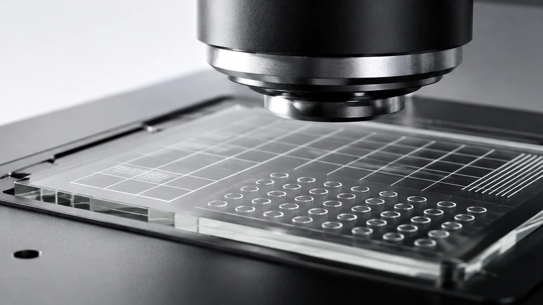
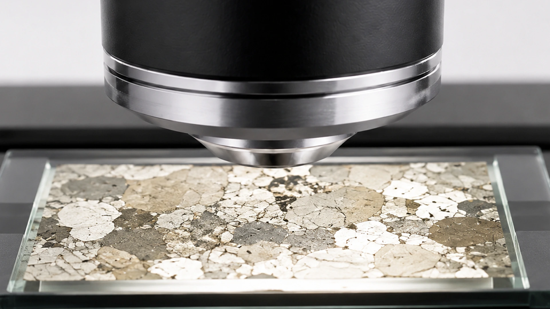
SLX uses a reflected-and-transmitted-light frame that switches flexibly between reflected and transmitted observation. Stage-position adjustment accommodates different sample heights, so metal workpieces, mineral and rock sections, glass, translucent materials and selected special samples can all be observed on the same platform.
04 / Configuration extension
Every precise operation, more controlled and stable.
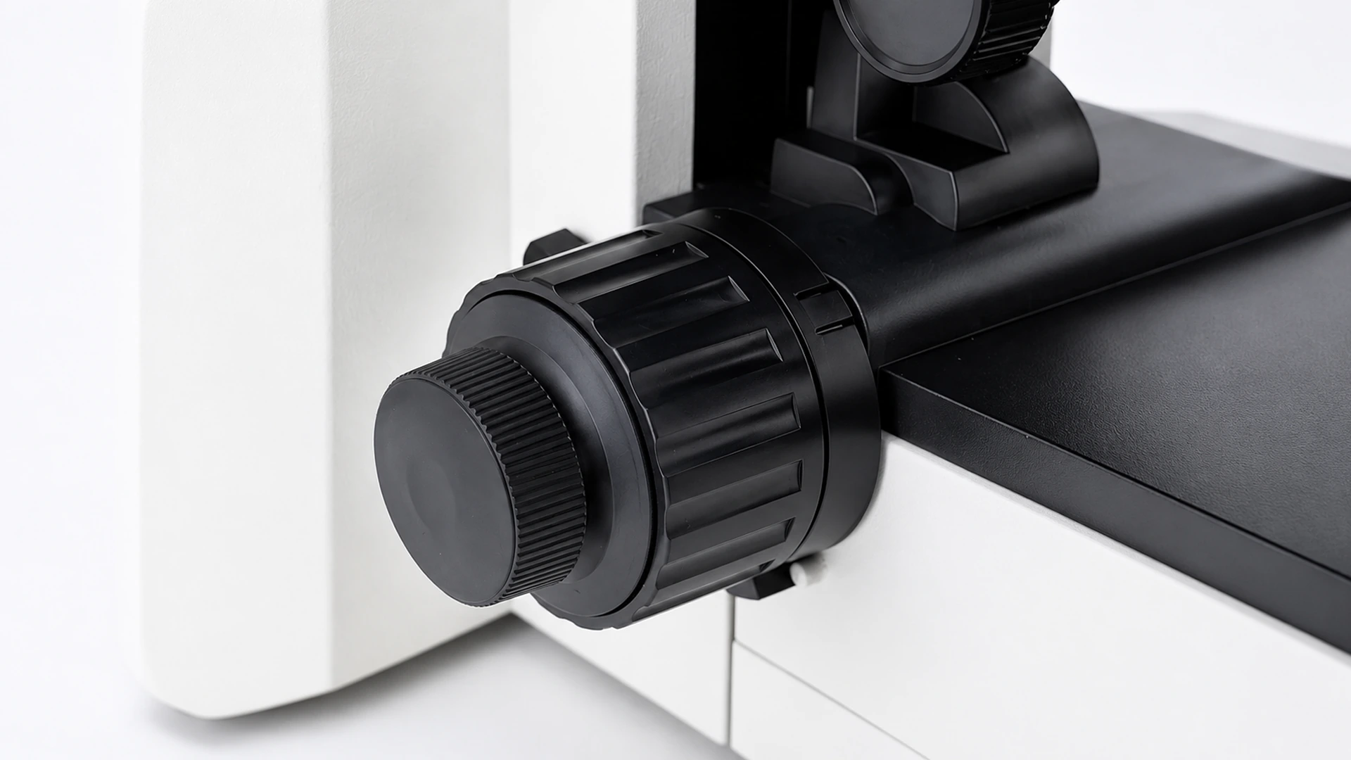
A double-layer mechanical stage provides low-position coaxial X and Y adjustment over 75 × 50 mm. Coaxial coarse and fine focus supports rapid positioning and precise adjustment, while an adjustable safety stop helps reduce accidental contact between the sample and objective.
Model Configuration
One platform. Three primary observation directions.
The SLX Series uses one microscopy platform with different primary measuring heads selected for the sample type and observation task. Specialist observation and analysis modules can be added for individual projects.
SLX-1000: bio-fluorescence configuration.Bio-fluorescence direction
SLX-1000
For fluorescence, biological samples and multimodal observation.
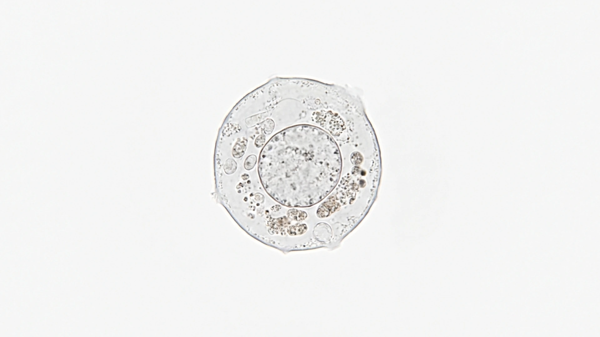
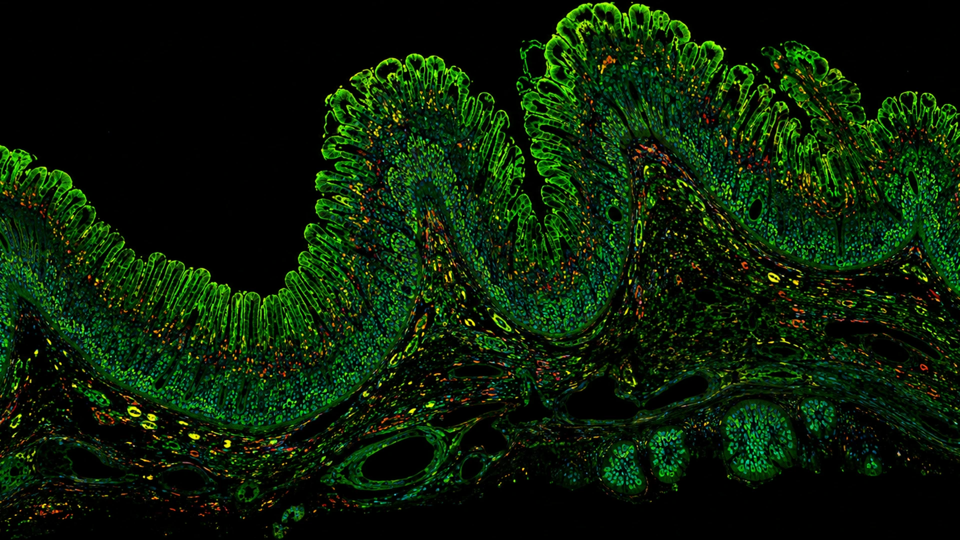
Built around bio-fluorescence observation, SLX-1000 is suited to microscopy of cells, tissue and related biological samples. The shared platform supports routine observation, fluorescence imaging and 3D measurement in one continuous workflow.
Fluorescence observation · multimodal observation · 3D measurement
SLX-1100: metallographic-analysis configuration.Metallographic-analysis direction
SLX-1100
For metal structures, material surfaces and industrial analysis.
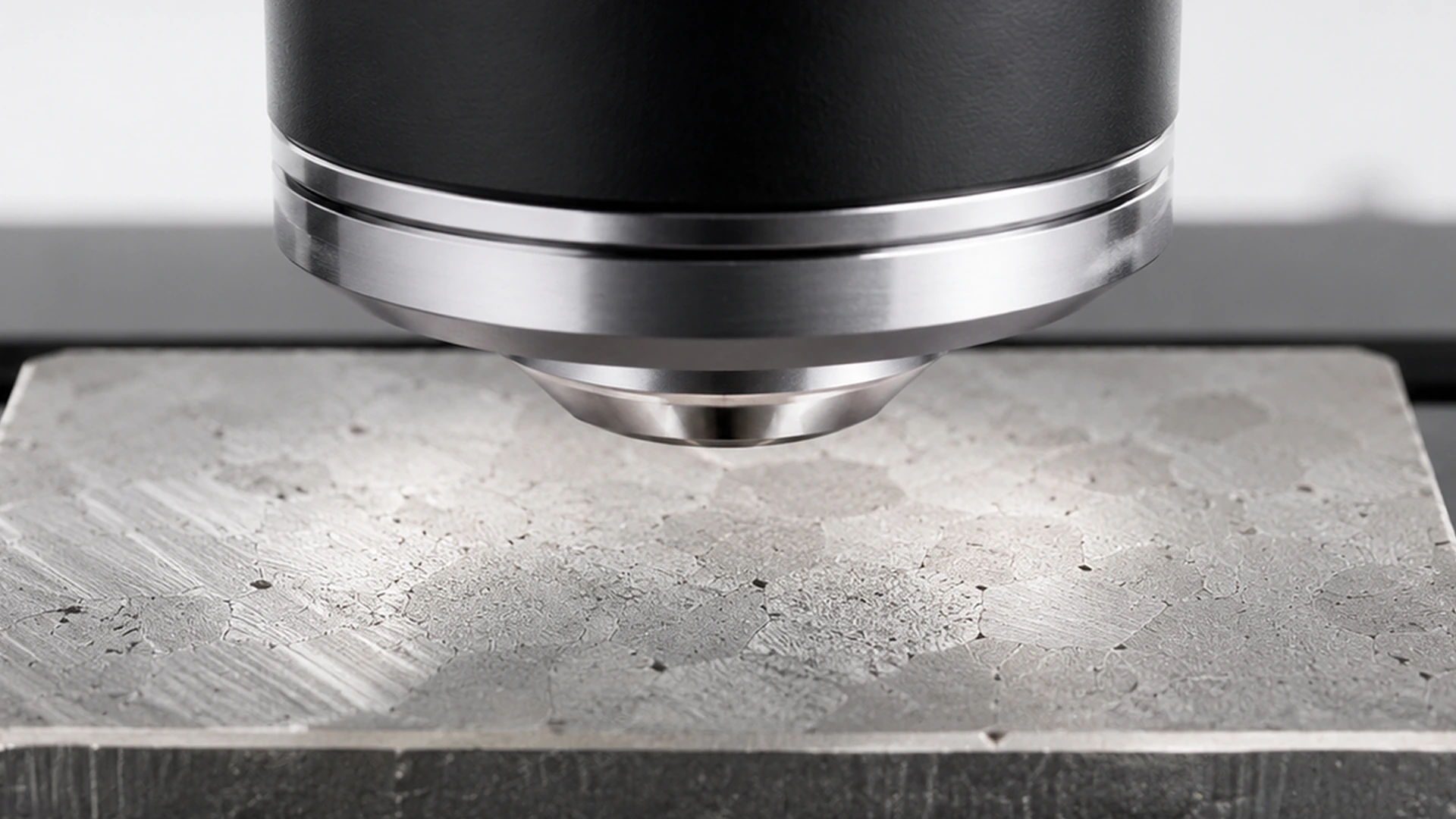
Built around metallographic and materials observation, SLX-1100 is suited to microscopic analysis of metal structures, machined surfaces, coatings and industrial samples. It supports overview observation, local detail and 3D measurement for materials research, quality inspection and failure analysis.
Metallographic observation · materials analysis · 3D measurement
SLX-1200: professional polarized-light configuration.Professional polarized-light direction
SLX-1200
For crystals, fibres and anisotropic materials.
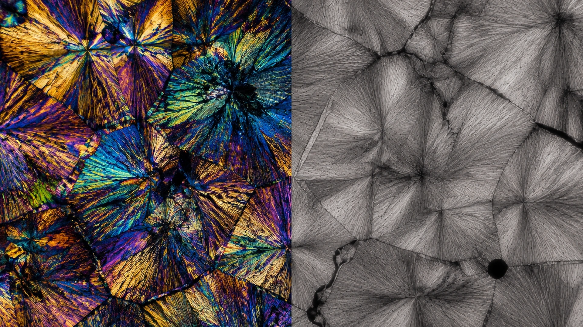
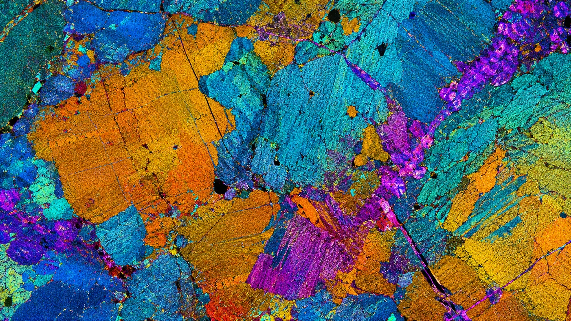
Built around professional polarized-light observation, SLX-1200 is suited to crystals, minerals, fibres and materials with directional characteristics. Polarized-light observation makes structural and directional differences easier to distinguish and supports further analysis with 3D measurement.
Polarized-light observation · orientation analysis · 3D measurement
Infrared, Raman and other specialist observation and analysis modules can be added for individual projects.
Technical Specifications
Complete Product Information
Only core specifications are shown here. Get the full product brochure for complete selection details.
Explore detailed specifications, model configurations, and additional product information.
Full Specifications
Model Configurations
Product Details
Core Specifications
The following shows the main models and core specifications.Swipe to view full specifications
Configuration Item
SLX-1000
SLX-1100
SLX-1200
Main direction
Bio-fluorescence and 3D measurement
Metallographic analysis and 3D measurement
Professional polarization and 3D measurement
Typical Samples
Fluorescent samples, biological samples and transparent samples
Metals, coatings, fractures and material structures
Crystals, fibers, minerals, films and anisotropic materials
Main Observation Focus
Fluorescence signals and multimode microscopic observation
Metallographic structures and industrial surface analysis
Material orientation, structural differences and polarization features
3D measurement
Supported by the main measurement heads; exact capability depends on configuration
Large field of view splicing
Supports sample overview and local review according to system configuration
2D measurement and result output
Shared capability of the series platform
Professional extensions
Infrared, Raman and other professional measuring heads / function modules are project options
Bio-fluorescence and 3D measurement direction
SLX-1000
Typical Samples
Fluorescent samples, biological samples and transparent samples
Observation Focus
Fluorescence signals and multimode microscopic observation
Analysis Direction
2D measurement, 3D measurement and result output
Metallographic analysis and 3D measurement direction
SLX-1100
Typical Samples
Metals, coatings, fractures and material structures
Observation Focus
Metallographic structures and industrial surface analysis
Analysis Direction
2D Measurement, 3D Measurement and Material Analysis
Professional polarization and 3D measurement direction
SLX-1200
Typical Samples
Crystals, fibers, minerals, films and anisotropic materials
Observation Focus
Material orientation, structural differences and polarization features
Analysis Direction
Two-dimensional measurement, three-dimensional measurement and polarization-assisted analysis
Shared platform capabilities
Shared configuration notes
3D measurement, large-field stitching, 2D measurement and result output are platform capabilities.
Infrared, Raman and other professional measuring heads / function modules are project options.
Specific objectives, illumination, cameras, measurement ranges and professional extension parameters are subject to the official brochure and project configuration.
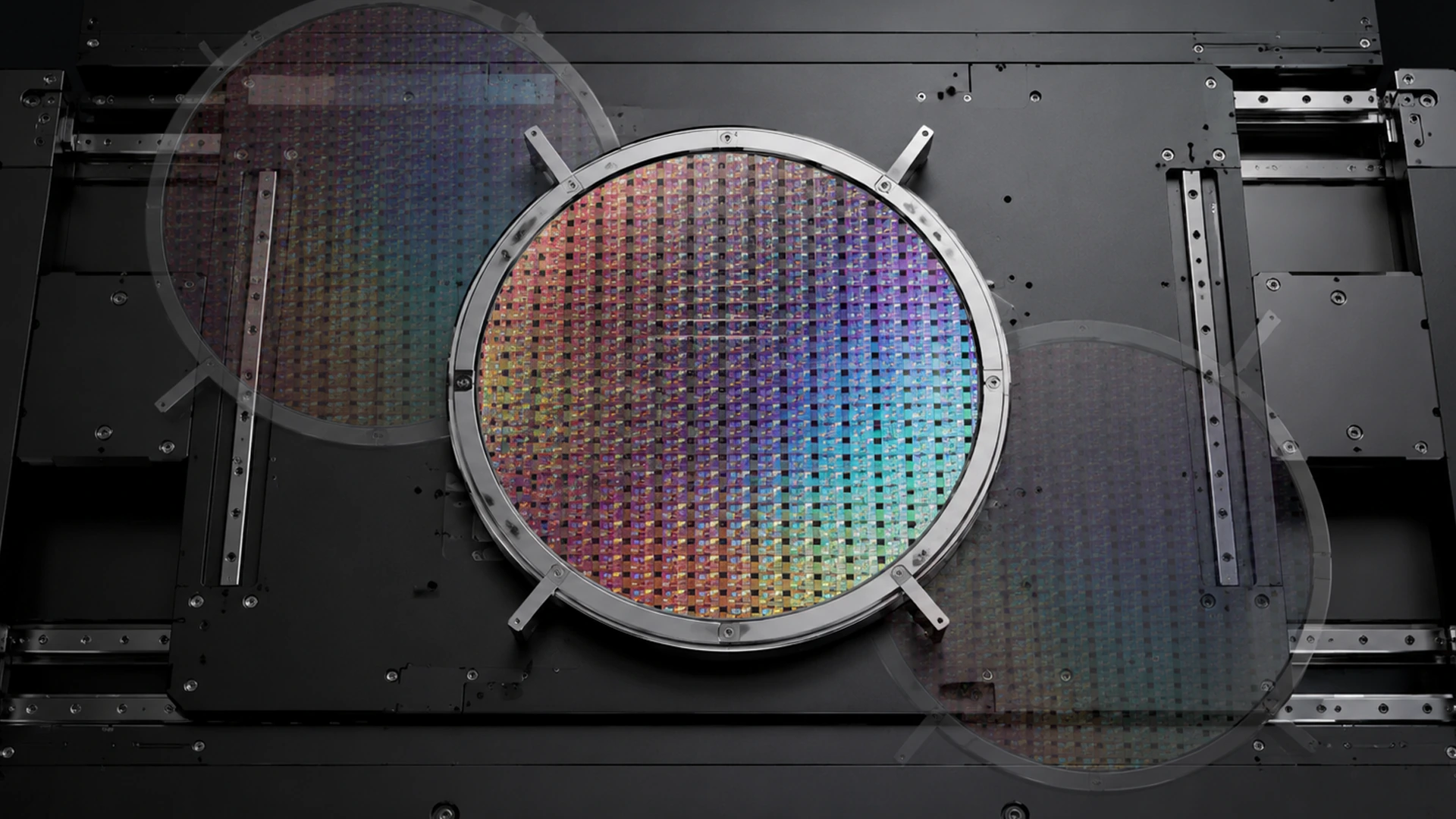
Multi-mode observation of wafer, chip, MEMS, package structure, solder joints, foreign objects and failure locations; carry out infrared internal observation and size and morphology analysis according to the configuration.
Metals and Materials Research
Observation and analysis of metallographic structure, grains, inclusions, fractures, coatings, corrosion, composite materials and surface structures.
Bio-Fluorescence and Life Science
Brightfield, fluorescence and correlation mode observation of fluorescent samples, cells, tissue sections, transparent samples and biological structures.
Polarization and Professional Micro-Area Analysis
Crystals, minerals, fibers, films, stress-related materials, as well as professional micro-area observation and analysis of Raman, infrared, etc. according to configuration.